Eye examination and consultation is test series performed to determine the quality of vision and field of view. This examination is also useful for diagnosing eye disorders and determine the treatment appropriately.
Generally, eye tests are recommended to be carried out routinely, even if there are no complaints, with the aim of detecting eye disorders early on. This is important, considering that eye disorders that are still in a mild stage can occur without causing symptoms that the sufferer is aware of.

Indications for Eye Examination and Consultation
How often eye examinations and consultations are performed generally depends on the age of the patient. The explanation is as follows:
Baby
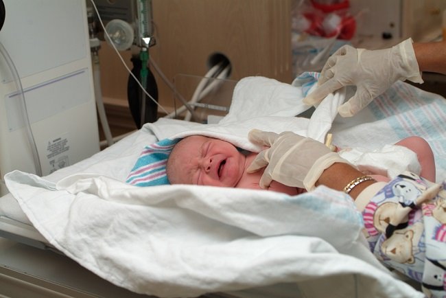
At birth, the baby's eyes should be checked to make sure there are no infections, birth defects, cataracts, glaucoma, and eye tumors. Further eye exams are recommended when the baby is 6–12 months old. The goals include checking the development of eye visual acuity, muscle movement, and eye coordination.
toddler
Eye examination in toddlers can be done when he is 3-5 years old. This is intended to prevent eye disorders that are prone to occur in toddlers, such as lazy eyes (amblyopia), Crossed eyes, and nearsightedness, can be detected early.
Children and teenagers
In this age range, nearsightedness is the most common eye problem, but is rarely noticed. Therefore, so that nearsightedness can be detected and treated early, children and adolescents are advised to have their eyes checked 1-2 times a year.
Mature
Eye examinations and consultations for adults with healthy eyes are recommended as follows:
- Ages 20–39: every 5–10 years
- Ages 40–54 years: every 2–4 years
- Ages 55–64 years: once every 1-3 years
- Ages 65 and over: every 1-2 years
Meanwhile, people with the following conditions require more frequent eye examinations and consultations:
- Using glasses or contact lenses
- Suffering from diabetes
- Suffering from high blood pressure (hypertension)
- Have a family history of glaucoma
- Regularly taking medications that affect eye health, such as corticosteroids, tamsulosin, birth control pills, cholesterol medications, antihistamines, diuretics, and antidepressants
Apart from being a routine health check, eye examinations and consultations are also recommended for people who experience the following symptoms:
- Red and sore eyes
- Blurred vision
- Double vision
- Sensitive to light
- There is a small object that floats in sight (floaters)
Eye Examination and Consultation Warning
The series of tests in the eye examination and consultation are painless and safe to perform. However, there are several things that patients need to know before undergoing an eye examination and consultation, namely:
- Tell your doctor if you are taking any medicines, supplements, or herbal products.
- Tell your doctor if you have eye problems or other ailments.
- Tell your doctor if you are allergic to eye drops.
Some eye examination procedures may involve administering eye drops that can interfere with vision for several hours. Therefore, it is advisable to invite relatives or family to accompany during and after the procedure.
Before Eye Examination and Consultation
Eye examination and consultation will be carried out by an ophthalmologist. There is no special preparation for this examination. However, patients are encouraged to prepare questions that they want to ask the doctor so that they can get as complete and complete information as possible.
In addition, patients who have previously used glasses or contact lenses are advised to take them along with their previous eyeglass prescription if available.
Eye Examination and Consultation Procedure
An eye exam and consultation usually lasts 45–90 minutes. The length of the eye examination depends on the method of examination carried out and the overall condition of the patient's eyes.
The eye examination begins with a consultation session. Patients are encouraged to inform the complaints they feel, whether related or not with the eyes. The ophthalmologist will also ask about the patient's and family's medical history, including a history of eye diseases, as well as any medications being used.
Next, the doctor will conduct a direct eye examination by observing the possibility of disturbances in the eyelids, eyelashes, and front eyeball.
After that, the examination can be continued with several series of tests, such as:
1. Visual acuity test
Visual acuity tests or eye vision tests are performed by displaying a chart containing letters of varying sizes, called snellen chart.
The patient will be positioned at a distance of 6 meters from the snellen chart, then asked to look at the same time to mention the letters designated by the doctor. If the results of the visual acuity test are abnormal, the doctor will perform a refraction test to determine the correct size of glasses or contact lenses.
2. Refraction test
Refraction tests are generally carried out using the method trial and error with tools such as glasses, you can use phoropter or trial lenses. When the patient wears phoropter or trial lens, the doctor will change the lens of this tool until the patient can clearly see the letters that were not visible before snellen chart.
With trial lenses, The doctor will also adjust the comfort of the lens being tested for daily use. The patient will be asked to walk, look around, or read, then assess whether the lens is suitable for him.
This test is useful for detecting refractive errors, such as nearsightedness (myopia), farsightedness (hypermetropia), old eyes (presbyopia), and cylinder eyes (astigmatism), as well as to determine a prescription for glasses or contact lenses.
3. Visual field test
The visual field test is useful for measuring how wide a person's eyes can see when compared to the normal eye area. The doctor will ask the patient to stare at an object that is located in the midline from the patient's front.
While viewing the object, the patient is asked to tell the doctor about another object moving sideways. How far the other object can still be seen by the eye, without moving the eyeball, from that the doctor assesses how wide a person's field of view is.
This visual field test is useful for measuring the range of vision that can decrease due to glaucoma or stroke.
4. Test slit lamp
Test slit lamp This is done using a device that shoots a thin line of light into the eye. With slit lamp, The doctor can see abnormalities in the eyelids, skin and tissue around the eyes, the surface of the eyeball (cornea and conjunctiva), the iris (iris), and the lens more clearly.
Sometimes, your doctor may give you eye drops to dilate the pupil, so that the deeper parts of the eye can be seen more clearly. This examination can detect eye lens abnormalities (cataract), retina (retinal detachment), and macular degeneration.
5. Tonometry
Tonometry uses an instrument called a tonometer to measure the pressure inside the eyeball. This test will help doctors in diagnosing glaucoma.
There are different types of tonometers. There are tonometers that are manually touched directly to the surface of the eyeball, some are digital machines and do not need to be in direct contact. If using a manual tonometer, the patient will be given anesthetic drops, so this procedure remains comfortable to undergo.
In addition to the tonometer, the eyeball pressure test can also be done using the doctor's finger by feeling the consistency of the patient's eyeball. However, this examination is subjective.
6. Ultrasound (USG) of the eye
Ultrasound of the eye uses sound waves to produce images of the structures inside the eye. This test is useful for evaluating eye tumors, cataracts, or bleeding in the retina.
7. Analysis cornea and retina
With certain machines, doctors can analyze abnormalities in the curvature of the cornea that can cause visual disturbances, such as astigmatism. This test is also useful for evaluating the shape of a patient's cornea before undergoing LASIK, receiving a corneal transplant, or choosing the right contact lens.
Apart from the cornea, the surface and all layers of the retina can also be mapped using a computer. This examination will make it easier for doctors to analyze retinal diseases that are difficult to examine with simpler examinations, such as: slit lamp or ophthalmoscope.
8. Fluorescein angiogram
This test is done by injecting a special dye (contrast) called fluorescein into the veins in the arm. This substance will move quickly to the blood vessels in the eye.
A special camera is used to photograph the flow of the substance in the blood vessels behind the eye. This test will make it easier for doctors to detect disorders of blood flow in the retina as well as abnormalities in the blood vessels in the eye.
Not all of the above examinations will be performed every eye consultation. The doctor will determine the examination needed by the patient based on the patient's age, complaints, and eye condition.
After Eye Examination and Consultation
After the examination, the doctor will inform the patient of the test results. From the results of these tests, the doctor will conclude several things to the patient, namely:
- Is there any disturbance that occurs in the patient's eye?
- Does the patient need to use vision aids or replace the lenses of glasses that have been used?
- Whether or not further treatment is needed, other than the use of visual aids
Side Effects of Eye Examination and Consultation
Side effects of eye examinations and consultations can occur if the doctor dilates the pupil (dilation) with eye drops to the patient. Side effects of dilatation itself generally only occur in the short term. Some of the side effects are:
- Sensitive to light
- Blurred vision
- Difficulty focusing when looking at close objects
- Stinging feeling when eye drops are inserted