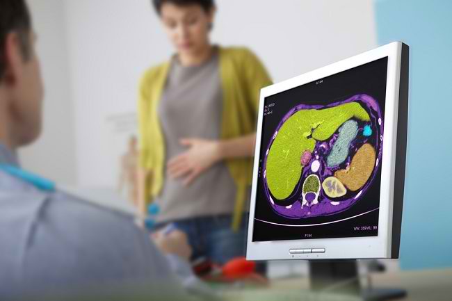
A cancer pathology report is a report on the results of a biopsy performed by a cancer patient or a patient suspected of having cancer. This pathology report plays an important role in helping doctors determine the diagnosis and treatment steps for cancer.
Pathology is a branch of medicine that studies the causes and processes of disease. Thanks to pathology, doctors can diagnose diseases more precisely, so that appropriate treatment can be given.

There are many branches of pathology. One of them is the pathology of cancer. Cancer pathology reports are made by anatomical pathology specialists (SpPA) who are in charge of examining tissue samples or patient body fluids in the laboratory.
How is a Cancer Pathology Report Created?
When suspecting the presence of cancer cells in the patient's body, the doctor will advise the patient to undergo a series of examinations, ranging from a physical examination to supporting examinations that include radiological examinations, such as X-rays, CT scans, MRIs, and ultrasounds.
In addition to these examinations, the doctor will also advise the patient to undergo an examination of samples of fluids or organs suspected of having cancer.
Sampling of fluid and tissue can be done by several methods, namely biopsy, aspiration (sucking it with a syringe), endoscopy, or surgery.
Samples of tissue and body fluids taken can be:
- Lumps in the body, for example in organs or lymph nodes.
- Lumps or abnormalities on the skin that are suspected of being cancer.
- Urine.
- Sputum.
- Vaginal discharge.
- Fluid around the spinal cord and brain (cerebrospinal fluid).
- Fluid in the abdominal cavity (peritoneum).
- Fluid in the chest cavity or lungs.
This sample is then sent to a pathology laboratory for processing and examination by a doctor. Usually, the analysis process takes 10-14 days. When finished, the pathology report will be sent back to the doctor who treated the patient.
Through cancer pathology reports, doctors and patients can get information about the appearance, shape, and size of the tissue and cells of the patient's body affected by cancer.
This cancer pathology report will greatly assist doctors in determining the diagnosis of cancer and its severity (stage of cancer). Once the diagnosis is confirmed, the doctor will provide further treatment.
What Information Is Included in the Cancer Pathology Report?
The following information is generally included in pathology reports:
1. Patient data
This information includes full name, gender, age and date of birth, medical history, and current diagnosis (if any). In addition, information regarding the type and date of the inspection was also provided.
2. General description of the tissue sample or fluid being examined
Comprehensive information about the weight, shape, and color of the patient's tissue or body fluids being examined.
3. Microscopic description
This information is a detailed explanation of the appearance, shape, and size of the patient's tissue cells as seen through examination with a microscope.
4. Final diagnosis
This information is the most important part in the cancer pathology report because it contains the conclusions of the results of the examination. If the diagnosis is a tumor, then this section will explain whether the tumor is benign or malignant (cancerous) and its size.
In addition, there is also information regarding the following 3 things:
- Stage of tumor/cancerThis information shows how heavy the cancer cells are growing and whether they have spread to other organs. Cancer cells that still resemble normal cells are classified as low-grade cancer cells. On the other hand, cells that look very different from normal cells are classified as moderate or severe cancer cells.
- Tumor/cancer marginWhen taking a sample on an area that is suspected of being cancerous, the doctor also takes a sample in a normal area that is around it. This sample is also known as a margin sample. The margin sample will be examined for the presence or absence of cancer in the normal area.
- Stage or stage of the tumorGenerally, anatomical pathologists will determine the stage of cancer based on the TNM classification, namely the size and location of the tumor (T), whether tumor cells have spread to nearby lymph nodes (N), and metastasis or whether the tumor has spread to other organs of the body (M). ).
5. Other inspection results
Anatomical pathologists may perform more specific tests or examinations to obtain more in-depth information about the tumor or cancer in the patient's body. The results of these additional tests or examinations will be described in this section.
Examples of these other tests could be genetic testing or special staining techniques on samples of tissue or fluid suspected of having cancer cells.
6. Synoptic report or summary
If the tumor or cancer has been removed, the anatomical pathologist will include a brief report in tabular form. This section is considered important in determining treatment options and the patient's chances of recovery.
7. Comment field
There are times when the results of the examination are not so clear that it is difficult to diagnose. Pathology specialists can use the comments field to provide recommendations for examinations or other tests to clarify results, if needed.
This column can also include other information that can help the doctor to determine the course of treatment and care for the patient.
8. Doctor and laboratory data
At the end, there is the name and signature of the anatomical pathologist, as well as the address of the examining laboratory.
Cancer pathology reports are technically written in medical language and may be difficult for patients to understand. However, the doctor who treats the patient will explain it in detail.
Patients are also advised to keep a copy of the report for themselves, so that one day the doctor can use it again when checking their medical history. A copy of the report can also be brought by the patient when he consulted another doctor if he wanted to ask for it second opinion.


