Osteopetrosis is a condition of abnormal bone density, which makes it easy to fracture. The condition is triggered by a disruption in the function of osteoclasts, a type of bone cell. Under normal circumstances, osteoclasts function to break down old bone tissue as new bone tissue grows. In people with osteopetrosis, osteoclasts do not destroy the bone tissue, causing abnormal bone growth.

Osteopetrosis is a disorder due to heredity, so it cannot be prevented. Routine screening for genetic disorders before delivery, followed by appropriate care and therapy, can help people with osteoporosis lead normal lives.
Symptoms of Osteopetrosis
Experts divide osteopetrosis into several types, each of which has various symptoms, namely:
- Autosomal dominant osteopetrosis (ADO).
ADO is mild osteopetrosis, which usually affects people aged 20-40 years. ADO is the most common type of osteopetrosis, and is estimated to occur in 1 in 20 thousand people.
Patients with osteopetrosis have a reduced risk in children by 50 percent. One gene mutation from either parent is enough to trigger ADO in a person.
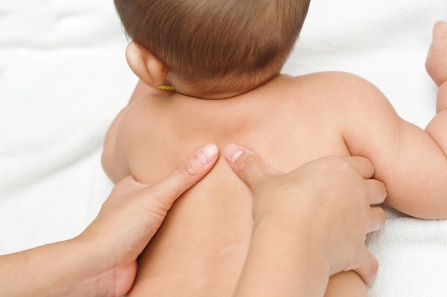
In some cases, ADO does not cause symptoms. But in some patients, ADO can cause a number of symptoms such as headaches, fractures in many places, bone infections (osteomyelitis), degenerative arthritis (osteoarthritis), and scoliosis or abnormal conditions in the spine.
- Autosomal recessive osteoporosisetrosis (ARO).
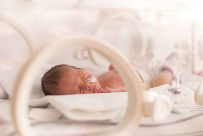
ARO is a severe form of osteopetrosis, which can affect a baby even when the baby is still in the womb. Babies with ARO have very fragile bones. Even during delivery, the baby's shoulder bones can be broken.
Until the age of one year, infants with ARO show symptoms of anemia and thrombocytopenia (lack of platelet blood cells). Other symptoms that can appear include facial muscle paralysis, hypocalcemia (low calcium levels), hearing loss, recurrent infections, slowed growth, short stature, abnormal teeth, and enlarged liver and spleen. In more severe cases, people with ARO can experience brain abnormalities, mental retardation, and frequent seizures.
A person can develop ARO if he inherits the mutated gene from each parent. However, parents who carry the gene may not have the disease.
ARO is rare, and only occurs in 1 in 250,000 people. However, this condition is very dangerous. If left untreated, the average child with ARO lives less than 10 years.
- Intermediate autosomal osteopetrosis (IAO).
IAO is a type of osteopetrosis that can be inherited from one or both parents. This type of osteopetrosis is also quite rare.
Although IAO is not as life-threatening as ARO, it can trigger a buildup of calcium in the brain. This condition can cause IAO sufferers to experience mental retardation.
- X-linked osteopetrosis.
This type of osteopetrosis is inherited through the X chromosome. Symptoms that appear in this type of osteopetrosis are immune system disorders that can lead to the development of more severe infections, as well as lymphedema. Other Symptoms X-linked osteoporosis is anhydrotic ectodermal dysplasia, which is a skin disease characterized by a lack of hair on the head and body, as well as a lack of the body's ability to produce sweat.
Causes of Osteopetrosis
Osteopetrosis is caused by mutations or changes in one of the genes involved in the development and function of osteoclasts, cells that play a role in the breakdown of bone.
Each type of osteopetrosis is caused by a different gene mutation, as described below:
- Mutations in the CLCN7 gene are known to be responsible for the majority of autosomal dominant osteopetrosis, 10-15% of cases autosomal recessive osteopetrosis, and a number of cases intermediate autosomal osteopetrosis.
- 50% of cases autosomal recessive caused by mutations in the TCIRG1 gene.
- X-linked osteoporosis caused by mutations in the IKBKG gene. This gene is located on the X chromosome. Chromosomes are parts of cells that contain genetic information, one of which is to regulate sex. In men who have one X chromosome, mutations in only one copy of the gene in each cell, will trigger this disorder. Whereas in women, who have two X chromosomes, mutations must occur in two copies of the gene. Therefore, X-linked osteoporosis generally occurs in men.
- In 30% of cases of osteopetrosis, the cause is unknown.
Diagnosis of Osteopetrosis
The diagnosis of osteopetrosis can be obtained through imaging tests, such as X-ray examinations. X-ray examination will help the doctor to see if there is an infection or fracture in the bone. Other imaging tests, such as a CT scan or MRI, may also be done as an investigation.
For patients with severe osteopetrosis, a blood sample will be taken for testing in the laboratory. In these conditions, calcium levels in the blood will be low, and levels of acid phosphatase and hormones calcitriol increased.
Osteopetrosis Treatment
Osteopetrosis in adult patients does not need to be treated, unless it causes complications. In this condition, the doctor will treat the fracture, or perform a joint replacement procedure.
On the other hand, osteopetrosis in infants should be treated immediately. Some methods for treating infants with osteopetrosis include:
- Giving vitamin D to stimulate osteoclast cells, so that the bone breakdown process can run normally.
- Giving calcitriol accompanied by restriction of calcium intake.
- Hormone therapy erythropoietin to treat anemia.
- Administration of corticosteroids to stimulate bone breakdown and treat anemia. This method may need to be used for several months or several years, and is not the method of choice for treating osteopetrosis.
- Surgery to treat fractures.
- Bone marrow transplant, to treat bone marrow disease and metabolic disorders. Although it is quite risky, the benefits derived from this method outweigh the risks.
Complications of Osteopetrosis
A number of complications that can be experienced by patients with osteopetrosis, among others, are bone marrow failure accompanied by severe anemia, bleeding, and infection. In addition, patients with osteopetrosis can also experience obstacles in the process of growth and development.


