Retinopathy of prematurity (ROP) is a congenital eye defect that often occurs in premature infants. ROP that is classified as mild can recover on its own as the baby ages. However, if severe, ROP can cause visual disturbances to blindness.
Basically, the blood vessels and retinal tissue of the fetus have begun to form and develop when the gestational age enters the 16th week. This part of the eye of the fetus will continue to develop until it can function properly after he is born at term (over 38 weeks).

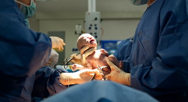
When a baby is born too soon or is born prematurely, the retina of the baby's eye is not fully developed, so it cannot function properly. This can interfere with his vision. This condition is called Retinopathy of Prematurity (ROP).
The sooner the baby is born, the higher the risk of getting it Retinopathy of Prematurity (ROP). This condition is also more at risk for twins born prematurely.
ReasonRetinopathy of Prematurity (ROP)
ROP is caused by the baby being born too soon, so the retina has not developed enough in the womb.
Until now, the exact cause of this is still not known with certainty. However, there are several risk factors that can make premature babies more susceptible to ROP, namely:
- Low birth weight
- Genetic disorders
- Fetal growth retardation (IUGR)
- Hypoxemia or lack of oxygen while in the womb.
- Infection in the uterus
Stages on Retinopathy of Prematurity (ROP)
ROP is divided into five stages, ranging from mild to severe. Here is the explanation:
Stage I
There was an abnormal growth of blood vessels in the retina, but still a little. Most infants with stage I ROP get better on their own without treatment with age. ROP stage I also generally does not interfere with vision.
Stage II
In stage II, found quite a lot of abnormal blood vessel growth around the retina. Just like stage I, infants with stage II ROP do not require treatment and as they age their vision will normalize.
Stage III
In stage III ROP, the abnormal blood vessels around the retina are so numerous that they cover the retina. This can interfere with the ability of the retina of the eye to support vision.
In some cases, infants with stage III ROP can improve without treatment and have normal vision. However, if the retinal blood vessels become enlarged and grow more and more, then treatment needs to be done to prevent retinal tears.
Stage IV
In ROP stage IV, the condition of the retina of the baby's eye is separated or partly torn from the eyeball. This is because the growth of abnormal blood vessels around the retina pulls the retina away from the wall of the eye. Infants with stage IV ROP should receive immediate treatment to prevent blindness.
V stadium
ROP stage V is the most severe condition in which the retina of the eye has detached from the eyeball completely. This condition must be treated immediately because it can cause visual impairment or permanent blindness if not treated immediately.
Treatment on Retinopathy of Prematurity (ROP)
Although stage I, stage II, and stage III ROP can recover as the baby gets older, this condition still needs to be checked and monitored regularly by an ophthalmologist.
This periodic examination is important so that the doctor can detect and evaluate the condition of the baby's eyes. If treated late or gets worse, ROP can cause the baby to develop various eye diseases, such as retinal detachment, nearsightedness, crossed eyes, lazy eye, and glaucoma, later in life.
Meanwhile, in advanced stages of ROP that are already severe, treatment needs to be done immediately to save the baby's sense of sight. Some of the steps for handling ROP include:
1. Laser therapy
Laser therapy is the most common treatment method used to treat ROP. This procedure aims to repair the periphery of the retina that lacks normal blood vessels. So that the retina looks clear and free from abnormal blood vessels that block it.
2. Cryotherapy
This treatment involves freezing the tissue around the retina to destroy the periphery of the retina to stop abnormal blood growth. The goal is the same as laser therapy for ROP.
3. Use of drugs
If needed, the doctor can give drugs that are injected into the baby's eyeball to stop the growth of abnormal blood vessels in the retina. This treatment method is generally done in conjunction with laser surgery.
4. Scleral Buckling
This treatment is used for severe cases of ROP. This procedure involves placing a flexible band of silicone around the eye circumference to encourage the torn retina to reattach to the wall of the eye.
5. Vitrectomy
This treatment is performed at stage V ROP. Vitrectomy is a surgical procedure in the eye to restore the position of the retina back to the eye wall.
ROP cannot be recognized by naked eye. The only way to detect and diagnose ROP is through an eye exam for ROP screening, which is performed by an ophthalmologist.
ROP screening will usually be done if the baby is born prematurely. If the results of the doctor's examination show that the baby has ROP, the doctor can determine further treatment steps to treat ROP according to the severity and condition of the baby.


