Cardiac examination is a procedure to detect disorders of the heart. In addition to diagnosing, heart examination can also measure a person's risk of developing heart disease before symptoms appear. In other words, this examination is also useful for preventing heart disease.
Heart disease is a disease that has a high risk of causing death. According to WHO, coronary heart disease ranks as the first cause of death in Indonesia, from the category of non-communicable diseases. Other data states, 15 out of every 1000 people in Indonesia suffer from heart disease.

Although very dangerous and can cause death, heart disease can be treated and also prevented. One way is to do a heart check to the doctor if you have symptoms of heart disease or have a risk for heart disease.
Cardiac examination itself consists of two types, namely non-invasive examination and invasive examination. Non-invasive tests such as ECG, echocardiography, stress test, Holter monitoring, and radiological examination. Invasive examinations include angiography and cardiac electrophysiology.
Types of Cardiac Examination
Cardiac examination is divided into several types, namely:
1. Electrocardiogram
An electrocardiogram (ECG) is a test to record the electrical activity of the heart. ECG is an examination that is often used to monitor heart conditions and detect heart problems quickly.
2. Echocardiography
Echocardiography is an examination of the heart using sound waves. Echocardiography is useful for monitoring the condition of the heart, including the condition of the valves and the ability of the heart to pump blood.
3. Pressure test(stress test)
Pressure test or stress test is an examination to determine the condition of the heart when the patient performs physical activities, such as running or cycling. This examination can be used to determine the presence or absence of disturbances in blood flow to and from the heart.
4. Holter monitoring
Holter monitoring is a test to monitor and record the electrical activity of the heart for 24 hours, with the help of a small device called a Holter monitor. Holter monitoring performed on patients who experience chest pain and heart rhythm disturbances.
5. Tilt-table test
Tilt-table test is an examination to find out what causes the patient to faint frequently. Tilt-table test can help doctors determine whether the cause of the patient's frequent fainting is related to blood pressure or heart rhythm disturbances.
6. Heart scan
A scan or imaging of the heart is done using radiology to get a general or specific picture of the heart, depending on the type of scan. The following are the types of examinations that include a heart scan:
- Chest X-ray
A chest X-ray is an examination that uses a beam of radiation to produce pictures of the internal organs of the chest, including the heart. This examination can be used to see the shape and size of the heart.
- CT scan of the heart
A heart CT scan is an X-ray examination that uses computer technology so that it can get images of the heart from various angles.
- Cardiac MRI
An MRI of the heart is performed using magnetic field technology and radio waves to produce images of the heart and the blood vessels around it.
7. Coronary angiography or cardiac catheterization
Coronary angiography or cardiac catheterization is an examination to detect and diagnose coronary heart disease and other heart conditions, such as heart valve abnormalities, heart function in pumping blood, pressure in the heart chambers, and oxygen levels in the heart.
8. Cardiac electrophysiology
Cardiac electrophysiology is an examination to map the electrical activity of the heart. This examination is performed on patients with heart rhythm disorders or arrhythmias. In some cases, doctors also use cardiac electrophysiology to measure a person's risk of sudden cardiac arrest.
Cardiac Examination Indications
Cardiac examination is carried out on people who have symptoms of heart disease, including:
- Chest pain or angina pectoris
- Easily tired or fainting easily
- Heart palpitations or irregular beats
- Hard to breathe
- Swelling in the legs
In addition, a heart examination can also be done to assess a person's risk of developing heart disease, especially in people who have the following risk factors:
- Suffering from high blood pressure
- Suffering from high cholesterol
- Suffering from diabetes
- Have excess weight
- Have a smoking habit
- Have an unhealthy diet
- Lack of exercise
- Have a family history of heart disease
- Experiencing heavy stress
Heart Check Alert
Cardiac examination is not recommended and is not even allowed in some conditions. Therefore, first consult with your doctor before you plan to undergo a heart examination.
During the consultation session, there are several things that the patient needs to do, namely:
- Tell your doctor about all current health conditions, including symptoms of heart disease that may be present.
- Tell your doctor about your past medical history, including symptoms of past heart disease and chronic conditions, such as high blood pressure, asthma, epilepsy, motor nerve disease, arthritis, and diabetes.
- Tell your doctor if you are allergic to contrast fluids and sedatives.
- Tell your doctor if you are pregnant or breastfeeding, especially before having a chest X-ray, CT scan, or MRI.
- Tell your doctor if you have tattoos, electronic devices, or metallic implants before having an MRI.
- Tell your doctor what medications you are taking, especially BETA blockers, such as bisoprolol and labetalol, isosorbide dinitrate, and nitroglycerin.
- Tell your doctor if you have a fear of tight spaces (claustrophobia).
Before Heart Check
The preparation that needs to be done before undergoing a cardiac examination can vary for each patient, depending on the type of examination to be carried out. But generally, doctors recommend the following things before the examination is carried out:
- Avoid drinking cold water or exercising before having an electrocardiogram (ECG), as this can affect the results of the test.
- Wear comfortable clothes and sports shoes before undergoing stress test.
- Do not eat for 4–8 hours before the CT scan. Also avoid consumption of drinks that contain caffeine.
- Remove all metal jewelry and body accessories before undergoing a chest X-ray, CT scan, or MRI.
- Do not smoke or drink caffeinated drinks for at least 2 hours before the carotid Doppler ultrasound. Also avoid wearing clothes or jewelry that cover the neck before undergoing this examination.
- Stop taking heart disease drugs, 24 hours before the heart examination is carried out.
Cardiac Examination Procedure
To get a diagnosis of a patient's heart condition, a cardiologist may run one or a series of tests. Before that, the doctor will ask about the symptoms experienced by the patient, as well as the medical history of the patient and his family.
The doctor will also check the patient's blood pressure and heart rate. If needed, the doctor will also perform blood tests to check complete cholesterol levels and C-reactive protein (CRP) levels. The results of these two tests can be used to assess the patient's risk of developing heart disease.
After that, the doctor will run a more specific heart examination. Here is the explanation:
Non-invasive examination
Noninvasive cardiac examination does not require making incisions in the patient's skin to insert medical devices. Types of cardiac examination with non-invasive methods include:
- ElectrocardiogramAn electrocardiogram (ECG) is performed by attaching 12–15 electrodes to the patient's body. These electrodes are connected to an EKG machine that will record the electrical activity of the patient's heart and print it on paper. The EKG procedure usually lasts about 10 minutes.
- Echocardiography
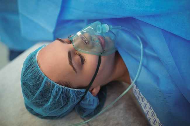
In transesophageal echocardiography, the scan is more complicated because the scanner must be inserted into the esophagus. The patient will be given an anesthetic during the examination. Therefore, the patient also cannot go home immediately and needs to be monitored first for several hours after the examination.
Echocardiography usually lasts less than 1 hour.
- Pressure test (stress test)On stress test, the doctor will ask the patient to walk treadmill or pedaling a stationary bike, starting at a low speed and gradually increasing. During physical activity, the patient is connected to an EKG machine and blood pressure.
stress test lasts about 15 minutes. During the examination, the doctor will monitor the patient's heart rhythm and blood pressure on a monitor. If the patient has symptoms, such as shortness of breath, chest pain, dizziness, or fatigue, tell the doctor.
- Holter monitoring
After 2 days, the doctor will compare the data from the Holter monitor with the records made by the patient, to diagnose the heart condition and the cause of the patient's complaints.
- Tilt-table testIn tilt table test, the patient will be asked to lie down on the examination table. Next, the table will be moved from a sleeping position to an upright or standing position. At the same time, the doctor will monitor the patient's heart rhythm, blood pressure, and oxygen levels. Usually, Tilt-table test lasts about 5–45 minutes.
- Chest X-ray
It is important to remember that the patient is required to hold their breath and not move during the shooting process, as movement can affect the resulting image.
A chest X-ray lasts quite a bit, only about 20 minutes.
- CT scan of the heart
After the patient enters the CT scan machine, detectors around the machine will capture images of the heart. During this process, the doctor will ask the patient not to move. The doctor will also several times ask the patient to hold his breath for a few seconds.
- Cardiac MRIIn an MRI exam, the patient will be placed on an examination table, which will be slowly pushed into the MRI machine, which is shaped like a tunnel. This MRI machine produces a noisy sound. Therefore, the doctor may give earplugs so that the patient is not noisy.
Once the patient is in the MRI machine, the doctor will give instructions through the microphone, so that the patient does not move and holds his breath while the image is being taken.
In some cases, the doctor will inject contrast fluid so that the images produced by the MRI examination are clearer and more detailed. Generally, an MRI scan lasts 30–90 minutes.
Inspectioninvasive
Cardiac examination with an invasive method is performed when a non-invasive cardiac examination does not provide a definite answer. In an invasive examination, the doctor will make an incision to insert the examination instrument into the body.
Some of the heart examinations with invasive methods are:
- Coronary angiographyCoronary angiography or cardiac catheterization is done by inserting a thin tube called a catheter into a vein in the arm or thigh. This catheter is then directed to the heart with the help of X-rays and contrast fluid, to produce an image of the coronary arteries of the heart.
- Cardiac electrophysiologyCardiac electrophysiology is performed by inserting electrodes into the heart through a catheter. The function of these electrodes is to send electrical signals to the heart and record the response from the heart.
After Heart Check
Patients can generally go home the same day after the cardiac examination. However, patients who are given anesthesia before the examination must first rest in the treatment room until their condition recovers, and ask their family or relatives to accompany them home.
For patients undergoing cardiac examinations with contrast agents, doctors will suggest drinking lots of water to speed up the removal of these fluids from the body.
Patients can find out the results of the cardiac examination on the same day or after several days, depending on the type of examination carried out.
On ECG, echocardiography, stress test, X-rays, and CT scans, the results can be known the same day. As for MRI, the results can only be known 1 week or more after the examination.
Depending on the results of the examination, the doctor may advise the patient to change a healthier lifestyle, undergo follow-up examinations, or provide medication.
Heart Check Side Effects
Cardiac examination is generally safe to undergo. However, in some cases, there are side effects that may arise from this procedure, including:
- Rash on the area of the skin where the electrodes were placed on the EKG or stress test
- Allergic reactions or kidney damage due to the use of contrast fluids
- Nausea, vomiting, and temporary low blood pressure, while undergoing tilt table test
- Heart rhythm disturbances and heart attacks after stress test, but this risk is very rare
- Infection, bruising, bleeding, or damage to the blood vessels at the catheter insertion site
- Blood clotting
- stroke